An anomaly scan also known as a Level 2 ultrasound or 20 week scan is routinely done for all expecting women between 18-20 weeks.
Why is an anomaly scan done?
An anomaly scan is an ultrasound done to assess the physical development of the fetus and to check the presence of malformations in the growing fetus. It is done between 18 and 20 weeks because before that the organs of the fetus cannot be delineated clearly on an ultrasound.
What does an anomaly scan detect?
The anomaly scan mostly looks at the head, brain, neck, spine, chest (heart and lung), abdomen (stomach bubble, kidneys and liver), face and arms and legs of the growing baby in the womb. The doctor performing the scan (sonographer) is specially trained for performing the scan.
The ultrasound also assesses location of the placenta, amniotic fluid around the baby and the length of the cervix.
Read: Posterior Placenta: 5 Myths (and Facts) You Need to Know
“The anomaly scan looks at certain features in the baby which are termed as soft markers of Down syndrome and looks at the blood flow to the uterus (uterine dopplers),” shares Dr. Anita Sabherwal Anand, Consultant Obstetrician – Gynecologist at Sitaram Bhartia Hospital in South Delhi.
It’s important to understand that not all congenital anomalies can be detected by ultrasound. It is relatively easy (90 to 95% accuracy) to identify problems with the structure of the head, brain, spine; locate a single kidney or an abnormal position of the kidney. However, it is often difficult to pick subtle abnormalities in the heart, abdomen and face of a baby.
“In certain conditions where for example, the expecting woman is obese or the placenta is anterior or there is too much or too little amniotic fluid then it can be difficult to assess the structure of the baby properly”, informs Dr Anita.
Why do I need an anomaly scan?
It is known that about 2-3% pregnancies will experience a problem with the growth and development of the baby. In most of these cases, visible defects can be picked up on an ultrasound.
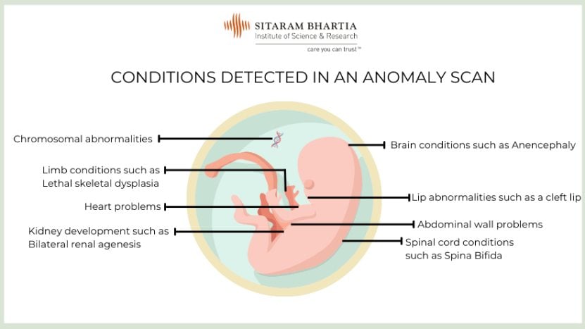
Some abnormalities, such as those in the skull and brain like anencephaly, or with the spinal cord like a large meningomyelocele or the absence of kidneys make it difficult for the baby to survive after birth.
Other abnormalities such as heart defects, tracheo-esophageal fistula or a cleft palate require early surgery to save the baby after birth.
“It is helpful to know the type of problem and whether it can be treated so that you and your doctor can understand and plan the medical care that is best suited for your baby.”
“However, expecting couples should be aware that while some abnormalities are easy to see at 20 weeks, there are others like those of the heart and intestines that become visible only after 28 weeks. Most importantly not 100% defects can be picked up by even the best of sonographers,” clarifies Dr Anita.
“You need to know that the absence of any structural abnormality does not rule out functional problems in the baby at birth.”
How is the anomaly scan done?
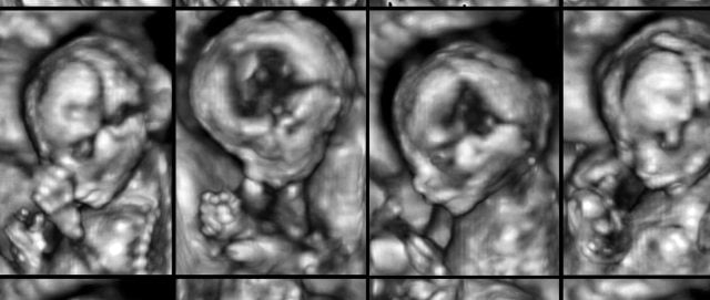
When you arrive for your ultrasound, the sonographer will ask you to lie on a table and uncover your abdomen. The ultrasound is usually performed in a dimly lit room so that the sonographer is able to get clear images on the screen.
The sonographer will apply gel over your belly and then place a probe on it. As the probe is moved over your belly, images of the fetus appear on the screen.
You may need to come with a full bladder so that the sonographer has a clear view of the uterus.
The various parts and organs of the baby are then evaluated in a sequence.
The sonographer will also take certain measurements such as:
- Head circumference (HC), which is the length around the baby’s head
- Abdominal circumference (AC), which is the length around the baby’s abdomen
- Femur length (FL), which measures the length of the thigh bone (femur)
These help in ascertaining the baby’s weight and growth in relation to the actual gestational age.
In some situations particularly if your baby is moving too much or is not facing the ultrasound probe, you may be required to come back in a couple of hours for re-evaluation.
If the placenta seems low lying, you may need to come with a full bladder so that the sonographer has a clear view of the uterus.
You will be informed if you have a low lying placenta, which means the placenta is positioned in the lower part of the uterus, toward the cervix. A repeat scan will be needed in the third trimester to determine whether the placental position has changed.
The ultrasound can help identify certain features in the baby that increase the risk of possibility of Down Syndrome. In such situations further testing in the form of amniocentesis (procedure of removing a few milliliters of amniotic fluid and sending it for genetic testing) may be required to confirm the diagnosis.
Does anomaly scan tell gender?
The gender of the baby can be detected in the ultrasound. However, prenatal sex determination in India is strictly prohibited by law.
Which week is the best for anomaly scan?
You can have an anomaly scan any time between 18-20 weeks, as there is no ‘best’ time for it.
In India, the Medical Termination of Pregnancy (MTP) Act does not allow terminations of pregnancy after 20 weeks. This is why the level II scan is offered at 18 to 20 weeks so that in certain circumstances, where severe abnormalities are detected and if the parents decide, they can be offered an MTP safely.
Is anomaly scan painful?
The anomaly scan, like any other ultrasound, is not painful.
“The sonographer may have to press down at times to get a clearer view, and this may be a little uncomfortable,” says Dr. Anita.
How long does the scan take?
Most of the time, the anomaly scan takes about 30 minutes. In very few cases, if your baby is moving too much or is not facing the ultrasound probe it may take a couple of hours or very rarely you may be called the next day to complete the scan.
What should I do if a condition is detected in my Level 2 ultrasound?
If a condition is suspected or identified during the scan, the sonographer will get a second opinion from a colleague.
“The sonographer will inform you if an anomaly is seen. Your doctor may advise another ultrasound or tests for further evaluation. Your doctor will explain what it entails and whether any treatment is available for it, “ says Dr. Anita.
“We may also refer you to a fetal specialist if required. We will guide you at every step, ensure you have all the information and support, not just from our maternity team but also our pediatricians and pediatric super specialists, should your baby require them.”
When will I get my anomaly scan report?
The sonographer will inform you of the results of the anomaly scan.
The anomaly scan report can be given as a draft, after the ultrasound is completed, for your doctor to review. The final ultrasound report is a typed document, and in a busy clinic, it is often made available after 2-3 hours or the next morning.
It will state any significant findings, the baby’s measurements, the amniotic fluid volume and the placental position.
“Most of the time, babies will grow as expected. In the event of an unexpected development, our entire team is here to support and guide you in making an informed decision that is best for you and your baby,” assures Dr. Anita.
Come in for a consultation Please Chat with us on WhatsApp to schedule an appointment.


Contributed and Medically Reviewed by Dr. Anita Sabherwal Anand
Senior Consultant, Obstetrician – Gynecologist
20+ years of experience
